Our brains have to do a lot of work when we watch a movie. There are plots to follow, dialogue to interpret, visuals to take in, and more. Now, scientists have created a detailed map of how the human brain functions during the process. Using data from functional magnetic resonance imaging (fMRI), a team from Massachusetts Institute of Technology mapped what different brain networks activate when subjects watch clips from a range of movies. They also saw how different executive networks in the brains are prioritized when watching easy versus difficult scenes. The findings are described in a study published November 6 in the Cell Press journal Neuron.
Inside the brain, different areas are interconnected. These various connections form functional networks that relate to how we perceive the world around us and behave. The majority of studies on the brain’s functional networks have been based on fMRI scans of people at rest. However, many parts of the brain or cortex are not fully active if external simulations aren’t present.
[Related: Why do jump scares terrify us so much? Blame evolution.]
In the new study, the team wanted to investigate whether screening movies during fMRI scanning could give any insight into how the brain’s functional networks respond to a series of complex audio and visual stimuli.
“With resting-state fMRI, there is no stimulus—people are just thinking internally, so you don’t know what has activated these networks,” study co-author and MIT neuroscientist Reza Rajimehr said in a statement. “But with our movie stimulus, we can go back and figure out how different brain networks are responding to different aspects of the movie.”
To map our brain on movies, the team used a previously collected fMRI dataset from the Human Connectome Project. The data included whole brain scans from 176 young adults that were taken while they watched a total of 60 minutes’ worth of short clips. The scenes ranged from independent movies including Two Men and Welcome to Bridgeville and larger Hollywood juggernauts like Home Alone, Inception, and The Empire Strikes Back.
The team averaged the brain activity across all of the participants and used AI to group together and pinpoint the brain networks that were activated, specifically within the cerebral cortex. The cerebral cortex is the brain’s outermost layer and is involved in many of the higher functions of the human brain including memory, learning, reasoning, problem-solving, and emotions..
Next, they examined how the activity within these different networks related what was in each scene, including people, animals, objects, music, speech, and narrative.
(D) The bar plot shows the percent signal change values for dynamic and static stimuli of six action categories in the action perception and social cognition clusters. The percent signal change values were computed based on the contrast of each stimulus condition vs. fixation. For the social cognition cluster, only vertices of the right hemisphere were included in the analysis due to a strong hemispheric lateralization of this cluster. Error bars indicate one standard error of the mean across subjects. (E) A cluster labeled as attention and eye-movement network.
(F) Dorsal attention network from Yeo’s 7-network parcellation. In (C) and (F), borders of relevant clusters are shown. CREDIT: Rajimehr et. al. 2024
They found 24 different brain networks were associated with specific aspects of sensory or cognitive processing, including recognizing human faces or bodies, movement, places and landmarks, social interactions, and inanimate objects, and speech.
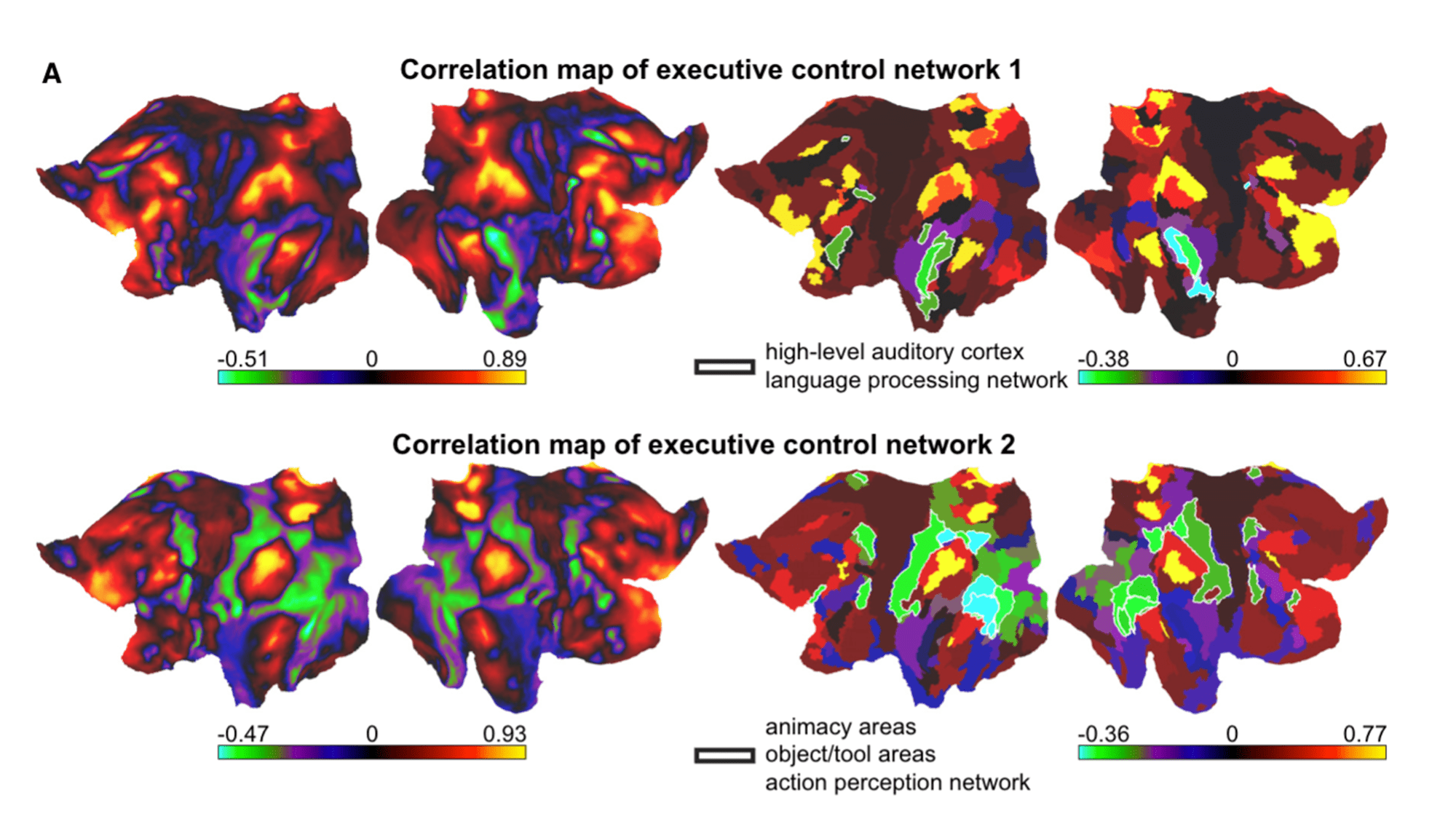
The scans also showed an inverse relationship between the brain regions that enable people to plan, solve problems, and prioritize information–-called executive control domains–and the regions of the brain with more specific functions.
When the film’s content was difficult to follow or more ambiguous like during Inception, activity was heightened in executive control brain regions. However, during more easy to understand scenes, the brain regions with specific functions–such as language processing–were the most dominated.
[Related: See the most detailed map of human brain matter ever created.]
“Executive control domains are usually active in difficult tasks when the cognitive load is high,” says Rajimehr. “It looks like when the movie scenes are quite easily comprehendible, for example if there’s a clear conversation going on, the language areas are active.”
However, during a complex scene with a lot of context, complicated wording, and ambiguity about what is going on, more cognitive effort is required.
“So the brain switches over to using general executive control domains,” said Rajimehr.
According to the team, figure research could investigate how brain network function differs between individuals of different ages or developmental or psychiatric disorders.
“Now, we’re studying in more depth how specific content in each movie frame drives these networks—for example, the semantic and social context, or the relationship between people and the background scene,” says Rajimehr.

